Main Joints Of The Human Body
Main joints of the human body- There are different types of joints in each part of our body like the cranial joints, Joints of vertebral, Joints of ribs and sternum, Joints of pectoral gridle, Joints of arm and forearm, Joints of hand, Joints of the pelvis, and Joints of the foot, etc bones have an important place in the composition of the human body. The structure of our body is formed by the bones, from which the body gets proper shape.
In the composition of the body, there are many types of bones such as small-large, long-thin, flat-round, etc. Bones together with muscles provide strength to the body. Bones join together to form many joints, some of which are movable and some are immovable. The ability to move the body is obtained by moving joints. Wherever two bones or cartilages meet together in the body, that joint is formed there. . Which helps us a lot in studying the human body.
JOINTS OF SKULL
Temporomandibular Joints
Temporomandibular joints Only this joint of the face is movable, it is the synovial joint, which consists of hind and gliding structures.
Atlanto Occipital Joint
There is a joint between the atlas and the occipital bone. It has hinged joints. In which the head is shaken [saying YES]
Main joints of the human body
JOINTS OF PECTORAL GIRDLE
Sternoclavicular Joint
It consists of the sternal end of the clavicle, the manubrium of the sternum, and the costal cartilage of the first rib. It is a gliding joint with elevation, depression, protection, and retraction motions.
Acromioclavicular Joint
It is a joint between the acromial end of the clavicle and the medial surface of the acromion of the scapula. It is a synovial joint and also has sliding motions. That helps in the free movement of the humerus bone on the shoulder joint.
Coracoclavicular Joint
It is a joint in the fibrous between the clavicle and the coracoids zone of the scapula. It is an immovable joint, which prevents the clavicle from separating from the scapula.
JOINTS OF THE VERTEBRAL COLUMN
In all vertebrae, there are joints from the second cervical vertebra to the sacrum. There is a cartilaginous joint between the bodies of the vertebra and a synovial joint is found between the vertebral arches.
JOINTS OF RIBS AND STERNUM
Costovertebral Joints
These are the synovial joints between the articular facets and tubercles on the tops of the ribs and the costal facets on the transverse segments of the thoracic vertebrae. They have gliding and rotating motions.
Sternocostal Joints
The ends of the costal cartilages of the ribs connect to the pits located on the lateral side of the sternum, forming the sternocostal joints. The joint of the first rib is immovable. Gliding motion occurs in the joints of the second to seventh ribs. These are synovial joints.
Interchondral Joints
The joints of the costal cartilages of the fifth to ninth ribs are called interchondral joints. They have gliding motion, which maintains adjustment during breathing. These are also synovial joints.
Manubriosternal Joints
It is the junction between the manubrium of the sternum and its body. This joint is a cartilaginous joint. There is a slight movement possible in this.
Xiphisternal Joints
It is a cartilaginous joint between the xiphoid region of the sternum and its body, in which there is also a slight movement.
Main joints of the human body
JOINTS OF HAND
Carpal Joints
These are the joints between the carpal bones of the proximal and distal rows. They have hinged joints. Because these bones are very close to each other, so there is a slight flexion and extension speed between them.
Thumb Carpometacarpal Joints
It is the joint between the trapezium and the proximal end of the first metacarpal bone. It is a saddle-type joint.
Metacarpophalangcal Joints
Common types of condyloid are formed between the apex of the metacarpal bone and the bases of adjacent phalanges. At these joints, the flexion, extension, abduction, adduction, and circumduction movements take place.
Interphalangeal Joints
Hinze-type joints are formed between the top of the phalange and the concave bases of the adjacent phalange. These joints have motions of flexion and diffusion.
JOINTS OF ARMS AND FOREARMS
Shoulder Joint [Glenohumeral Joint]
The shoulder joint or glenohumeral joint provides more movement than other joints of the body. This joint is of ball and socket type. This joint is formed by the spherical apex of the humerus in the form of a ball, fitting into the glenoid cavity of the scapula form socket. The articular surface is covered by fibrous tissue. The glenoid cavity is further deepened by a fibro-cartilaginous rim called the glenoid labrum, providing additional firmness without limiting movement and reducing the risk of dislocation. The scapula and humerus bones are held together by flexible capsules of ligaments, and the powerful muscles help maintain the bones’ position. The long tendon of the biceps muscle serves as the intracapsular ligament. It originates from the scapula just above the glenoid cavity, due to which the joint remains in the correct position
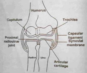
Radio Ulnar Joint
The elbow joint is a type of hinge joint. The movements of flexion and extension mainly take place in this joint. This joint is formed by joining the trochlea and capitulum, the trochlear notch of the ulna, and the head of the radius, located at the lower end of the humerus bone. The trochlea of the humerus fits into the trochlea notch of the ulna and joins the apex of the capitulum radius. In this joint, there are many ligaments that store strength outside the capsule.
This joint consists of two joints which are in the form of curved movable joints.
- The proximal radioulnar joint is located between the top of the radius bone and the radial notch of the ulna bone.
- The distal radioulnar joint is located between the top of the bone and the ulnar notch of the radius bone.
Both the joints have pronation and supination motions. In pronation motion, the palm moves down and in supination motion, the palm moves up. Around the apex of the radius are strong annular ligaments, which keep the contact of the radius and the radial notch of the ulna in order.
Radio Carpal Joint-
This joint is the joint between the lower vein of the radius and the carpal bones called scapholunate and triquetrum. Medial ligaments and radiocarpal ligaments are present outside the capsule of the joint.
In this joint, there is abduction, flexion, extension, adduction, hyperextension, and circumduction motions occur.
ARTICULATION JOINTS OF THE PELVIS
Sacroiliac Joint
There are two anterior and posterior joints between the sacrum and ileum bones; the anterior joint is the synovial joint, which has slight gliding and rotational motions that provide flexibility to the joint. And the posterior joint is a fibrous joint, which has a small movement and provides protection to the joint.
Symphysis Pubis
The filaments between the bodies of the pubic bones are fibrocartilaginous joints, which are almost immovable but have some movement during delivery.
Coxal And Hip Joint
It is a ball and socket type joint formed by the fit of the spherical end of the femur bone to the cup bone-deep acetabulum of the hipbone. The joint forming surfaces are covered with the articular cartilage. The acetabular labrum around the edges of the acetabulum is fibrous cartilage, through which the depth of the acetabulum cavity is further increased, in which the top of the femur is well adjusted.
The capsular ligament covers most of the femur’s cervix. The ligament on top of the femur starts from a small and coarse pits fovea at the apex and runs to the acetabulum. The synovial membrane covers both sides of the acetabular labrum and forms a shell around the ligament at the top of the femur. Several ligaments surround the joint capsule and give it strength; one of the most powerful is the iliofemoral ligament, which lies in front of the joint and allows the spread of the hip not far from the midline of the torso. It is possible to have flexion, diffusion, refraction, and elevation, medial and lateral rotation, and circumduction movements at the hip joint.
Knee Or Tibiao-Femoral Joint
The knee joint is the largest and most complex joint of the body. It is a synovial joint of the Hinze structure.
The knee joint comprises three synovial joints, one between the femur and the medial condyloid of the tibia, the other joint between the lateral condyloid of the bone, and the third joint between the patella and femur. The anterior part of the articular capsule is made of the tendon of the quadriceps femoris muscle, which also supports the patella. The articular capsule is called the Brach ligament, tibial or medial collateral ligament, and the fibular or lateral collateral ligament that extends to the femoral cortex, which gives it strength.
Within the joint, there are two anterior and posterior cruciate ligaments similar to a strong lobe crossing each other in the middle, extending from the intercondylar notch of the femur to the intercondylar eminence of the tibia and covered by the synovial membrane. They help to keep the joint stable and control the movement of the knee.
Within the joint, there are two semilunar discs of white fibrous cartilage, called menisci, on the crest of the articular conidia of the tibia. These are wedge-shaped, and their outer edge is thicker. It aids in stabilizing the joint by preventing lateral displacement of the bones and makes them thicker to fit the femoral condylar surfaces on the articular surfaces of the tibia.
Ankle Or Talocrural Joint
It is a hinge-type joint between the lower end of the tibia and its medial malleolus and the distal end of the fibula or the lateral malleolus and the upper surface of the talus. The joints get strength from deltoid and anterior, posterior, medial, and lateral ligaments. It has the speed of flexion and diffusion, but usually, flexion is called dorsiflexion, and variance is called plantar flexion.
Main joints of the human body
ARTICULATION JOINTS OF THE FOOT
Tarsal Joints
The tarsal bones are joined together by the dorsal, plantar, and interosseous ligaments. Gliding joints are sliding between them. The tarsal bone has a posterior joint or subtalar joint between the talus and the calcaneus. An anterior joint or talocalcaneonavicular joint formed between the talus and the calcaneus and between the talus and navicular bones. A transverse tarsal joint is formed between the calcaneus and the cuboids and between the talus and navicular bones. These three joints form a joint as a unit. Their axis of rotation includes a line, which is called the subtalar axis. There is the motion of inversion and aversion; that is, the foot rotates inwards and outwards.
Tarsometatarsal Joints
These are the joints between the four anterior tarsal bones and the metatarsal bone bases. These are gliding joints in which there is light movement.
Metatarsophalangeal Joints
These are the condyloid joint joints between the top and proximal phalanges bases of the five metatarsal bones.
Interphalangeal Joints
There is a hinge-type joint between the head of the phalanges and the concave bases of the connecting phalanges. These joints are retraction and diffusion movements.