STRUCTURE AND FUNCTION OF HEART
STRUCTURE AND FUNCTION OF HEART – The heart is the major and specialized organ of the blood circulation system. It is made up of cardiac muscles. Its size is the size of its own closed fist, on average, it is about 12 cm in length and 9 cm in width, it weighs about 250 grams to 390 grams in adult males and between 200 and 275 grams in adult females. The heart beats approx 72 times in a minute and 1 lakhs times in a day. The study of heart-related diseases is called cardiology. The hormone which stimulates the heartbeat of the heart is thyroxin. CARDIAC CYCLE plays important role in the functioning of the heart.
The structure of the heart is made more complicated because it is the mechanism that allows blood to be distributed throughout the body in sufficient amounts and return to the heart. This continuous process is accomplished with the help of two blood vessels: veins and arteries. The arteries that carry oxygen-rich blood from the heart and to other parts of the body are called arteries and the vessels which bring deoxygenated blood back to the heart are called veins.
LOCATION OF HEART
The heart is situated in the thoracic cavity between the lungs within the mediastinum. [Structure and function of heart] It is a muscular, hollow, contractile, conical organ that is located behind the sternum with a portion of the middle tissue of both lungs called the mediastinum, and some obliquely on the left side. Due to some being on the left side, one-third of it is situated from the midline of the body to the right side and two-third part is located on the left side.
The upper part of the heart is slightly wider, which is called the base, and is tilted to some right. The base is directed superiorly and slightly posterior. The lower point is called the apex which is slightly to the left and slightly forward and rests on the diaphragm. The APEX boundary reaches the intercostals between the fifth and sixth left ribs. Keeping a hand in this place gives the impression of a heartbeat.
It is important to know the location of the heart placing a stethoscope to hear the heart sound [lub-dub], placing an electrode on the chest to record an electrocardiogram [ECG], and performing cardiopulmonary resuscitation [CPR] depend on a knowledge of the heart position.

LAYERS OF HEART WALL
Basically, there are three layers of tissues in the heart wall. The outermost layers called PERICARDIUM, the middle layer of the heart wall is called MYOCARDIUM, and the inner layer is called ENDOCARDIUM.
PERICARDIUM
The pericardium is the outermost layer of the heart. The pericardium or cardiovascular is made up of two sacs. The outer sac is made up of fibrous tissues and is in continuity with the double layer of the serous membrane from the inside. In the external fibroid, the sac remains in continuity with tunica adventitia of the large heart vessels above and adjacent to the diaphragm at the bottom. Being fibrous and inelastic in nature, it prevents excessive dilation of the heart.[Structure and function of heart]
- The outer layer of the serous membrane covers the fibrous corpus of the parietal pericardium.
- The inner layer, the visceral pericardium or EPICARDIUM that is in continuation of the parietal pericardium, is affixed to the heart muscle.
The serous membrane consists of flat epithelial cells. It releases a serous fluid into the space between the visceral and parietal layers which prevents friction between the two layers during the heart pulse, which keeps the heart functioning freely.
MYOCARDIUM
The myocardium is the middle layer of the heart. The myocardium is made up of cardiac muscle fibers or cardiac myocytes. It contains special types of fibers which belong to the involuntary class. The thickness of the myocardium varies; it is thickest at the apex and becomes thinner towards the base. The left ventricle remains thicker because it has to do more work, in the same right ventricle, this layer is thinner because it only has to flow blood to the lungs. This layer is very thin in the atrium.
The myocardium is consist of three muscle fibers:-
- Fiber helps in the formation of the contractile unit of the heart
- fiber which helps in the formation of pacemaker
- fiber which helps in the formation of a conductive system of the heart
ENDOCARDIUM
The endocardium is the innermost layer of the human heart. The Endocardium is a thin, smooth, shimmering soft membrane within the heart. It is made up of flat epithelial cells that merge with the endothelium in the internal levels of the blood vessels by being continuous. The Endocardium is covered with all the four chambers and valves of the heart.
CHAMBERS OF THE HEART
The septum divides the heart lengthwise, and valves divide it crosswise. Each side of the heart thus has two chambers, one above the other. A thin membrane called the Endocardium lines each chamber. [Structure and function of heart] The entire right side of the heart deals with the exchange of impure blood and the left side deals with the exchange of pure blood. Both the right and left parts are again divided by a transverse septum, forming an upward and downward chamber. In this way, the entire internal cavity of the heart is divided into 4 chambers:
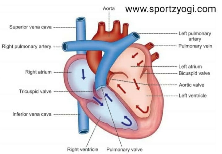
- Right atrium
- Right ventricle
- Left atrium
- Left ventricle
Both the atrium and ventricle on the left and the right side are connected by a hole. Valves are attached to these holes, the valves between the two chambers are in such a way that blood can only go from the atrium to the ventricle, but cannot return.
Right atrium
In this chamber, the deoxygenated blood returning from the body is stored in the entire body. Veins coming from different parts of the body are made up of two large veins, the superior vena cava [from the upper part of the body] and the inferior vena cava [from the lower part of the body], which come into the right atrium and opens separately. The right atrium only accepts blood, and the work of pumping blood has little to do, so its contraction is relatively low, that’s why its walls [membrane] relatively thin and weak. After receiving the blood, it has to push the blood so much that it opens the tricuspid valve and goes into the ventricle.
Right ventricle
Impure blood from the right atrium flows into the right ventricle via the right interventricular orifice. The contraction of the right atrium pushes blood, causing the tricuspid valve sutures to open themselves and impure blood to enter the ventricle. The second moment there is a contraction in the right ventricle, as a result of which the blood is pushed to drain out. The right ventricle has a hole called a pulmonary orifice.[Structure and function of heart]
Through this, the pulmonary artery emerges, which later goes out of the heart and divides into two parts and goes to the right and left lungs. As soon as there is a contraction in the right ventricle, impure blood goes through it to be purified into the lungs. As blood enters the pulmonary artery from the ventricle, the pulmonary valve between them closes and impure blood cannot return to the ventricle.
The ventricle walls are thicker and stronger than the right atrium because it has to be compressed more than the right atrium to pump blood so that it can reach the lungs to be purified.
Left atrium
The upper chamber of the left side of the heart is called the left atrium. It is smaller than the right atrium. Its walls are thicker than the right atrium. In this, two of the four pulmonary veins come from the right pleura [LUNGS] and two from the left pleura [LUNGS] through four holes and bring pure oxygenated blood to the left atrium.
Left ventricle
The lower chamber of the left part of the heart is the largest chamber and its reefs are also thicker than the reefs of all other chambers. It has a hole called an aortic orifice. Through this, the aorta carries blood to different parts of the body.
As a result of the contraction of the left atrium, purified blood fills the left ventricle. The second moment comes to the contraction of the left ventricle. Before this, the mitral valve between the left atrium and the ventricle is closed. On narrowing of the left ventricle, the purified blood opens the aorta’s door with a push and blood comes out of it. There are also valves on the aortic mouth that prevent the blood flow from returning. In this way, the left ventricle performs the most important function of providing nutrients and oxygen to the tissues in the body and also carrying the blood to the body.
Structure and function of the heart
VALVES OF THE HEART
There are valves in the heart to prevent the backward flow of blood.
- TRICUSPID VALVE: positioned between the right atrium and right ventricle
- PULMONARY VALVE: positioned between the right ventricle and pulmonary artery
- MITRAL VALVE: positioned between the left atrium and the left ventricle
- AORTIC VALVE: positioned between the left ventricle and aorta
STRUCTURE AND FUNCTION OF HEART
Pumping blood to the lungs
Blood from the body that enters the right side of the heart contains carbon dioxide, a gaseous waste the cells produced in generating energy. Blood comes in the right Atrium through the superior-inferior Vena cava. The Atrium fills the blood and then contract, forcing the blood through the tricuspid valve into the right ventricle.
After the ventricle is filled, the pressure pushes the tricuspid and valve to close, and the pulmonary valve, leading to the Pulmonary artery, to open, the ventricle contracts and the blood through the Pulmonary artery and into the lungs. In the lungs, through the exchange of gases carbon dioxide is removed from the blood, and oxygen is added in the blood. The oxygenated blood then flows through the pulmonary veins to the left side of the heart.
Pumping blood throughout the body
Oxygenated blood from the lungs enters and fills the Atrium. The Atrium then contracts which squeezes the blood through the mitral/ bicuspid valve into the left ventricle, after blood fills the ventricle, the mitral valve closes and the aortic valve opens. Blood pours into the aorta and flows through arteries to the body tissues.
Regulating blood pressure
Blood in the circulatory system like water in the pipes of the water system is always under pressure. Blood pressure refers to the force with which the blood pushes against the walls of the arteries. That force drives blood from the heart to all parts of the body. Structure and function of heart- Each person’s blood pressure reflects the amount of blood in the body, the strength and rate of the heart contraction, and the elasticity of the arteries.
As the heart pump in cycles, pressure in the arteries Rises and Falls during systole and diastole. Contraction of the heart produces systolic blood pressure, and relaxation produced diastolic blood pressure. The heart helps regulate blood pressure by producing a hormone that helps the kidney in eliminating Salt from the body. Excess salt may contribute to high blood pressure, which is called hypertension. Hypertension can injure the heart, brain, and Kidneys.
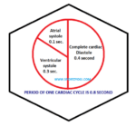
Regulating the heart rate
Both sides of the heart pump blood at the same time. As the right ventricle contract and send blood to the lungs, the left ventricle contract and screws blood out to the body. The heart cycle of activity has two periods, systolic and diastolic. Systolic occurs when the ventricle contract, and diastole when they relax. One complete contraction and relaxation of the heart muscle make up one heartbeat.
Structure and function of heart
READ MORE ABOUT- Structure and function of skin